Bright-field and dark-field illumination
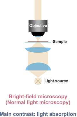
In normal bright-field microscopy, light is focused by an objective lens (characterized by its magnification and numerical aperture (NA), e.g. 50x/NA=0.75) onto the sample and the light either reflected (reflected light microscopy) or transmitted (transmitted light microscopy) by the sample is collected. The image contrast is primarily based on light absorption.


In dark-field microscopy, the inner part of the illuminating light beam is blocked by a mask. Thus, only a sheet of light is focused onto the sample. In other words, the sample is illuminated under an angle that is defined by the NA of the objective lens. Only the fraction of light is detected, which has changed its angle upon interaction with the sample. The image contrast is primarily based on light scattering, enhancing the contrast of, e.g., small particles and sharp edges.
